You Saw It Here First.
Here is the first published illustration that I have done in over 25 years:
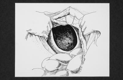
It will appear in the Journal of the American Veterinary Medical Association. What are you looking at? Well, it's a picture of what you would see if you had a horse being operated on for Morgagni's hernia. This is when there is a congenital hole in the diaphragm, usually on the right side, allowing the transverse colon of the horse to occupy thoracic space along with the heart and the lungs. Interestingly, this can happen not only with horses but also other animals and people as well, and the depiction of this situation in the horse was why the article was being published. So the rim of the hole is the diaphragm, and what's visible inside it is the hernial sac. Sometimes the sac is removed, but because it's so close to the pericardium (the membrane enclosing the heart), surgeons frequently decide to leave it there. The lumpy thing in foront of the hole is the colon that has been removed from that place. It was placed inside the abdomen where it belongs. The hole was closed up with surgical mesh and sutured in place. The dotted line is the location of the xiphoid process, which is a little 'peninsula' at the end of the sternum. The horse was lying on its back for the surgery.
I love this job. Wait'll you see the eyeball I just painted.
posted by craigellachie at 7:32 PM
![]()

4 Comments:
Eleanor! this is SOOOO COOOOOLLLL! way to go. I'm excited and proud of you.
Heidi
Wow! That's most impressive, Eleanor.
Keep sharing your drawings, please. We love seeing them.
Thanks, Anne and Heidi. I will post some more eventually, but they've got to be photgraphed yet.
Great galloping horse guts, that's good stuff!
~Valerie
Post a Comment
<< Home